


Guiding patient care in ambiguous melanocytic lesions
MyPath Melanoma is a gene expression profile (GEP) test that provides clarity in managing patients with ambiguous melanocytic lesions.
GEP provides objective information to clarify malignant potential
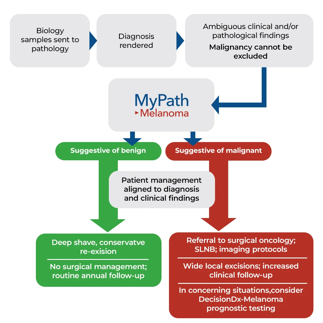
When patients are diagnosed with routine nevi or malignant melanoma, the treatment plans are clearly defined by medical guidelines. However, there is a lack of clarity and standardization in the most appropriate management plan for patients with ambiguous, atypical, and abnormal melanocytic lesions. MyPath Melanoma can clarify the underlying malignant potential of the lesion allowing providers to escalate or de-escalate surgical treatment and follow-up frequency.


Aiding dermatology providers in determining malignant potential
In recent studies and case reports, 1,2 ancillary diagnostic GEP testing has aided dermatologists in determining the malignant potential of these challenging melanocytic lesions. This allows providers to confidently align patient management to the malignant potential as determined by GEP.
MyPath Melanoma GEP testing provides an objective assessment of the expression levels of 23 genes and applies a proprietary algorithm to classify the lesion as suggestive of benign or suggestive of malignant.3 Numerous studies have demonstrated high sensitivity and specificity 4-8 and clinical utility 9–12 of GEP testing for dermatology providers.

Hear how MyPath Melanoma can be used in your practice
“When we need to make a decision on excision margins and future surveillance then the MyPath test has become a useful tool for me and my patients.”
Learn how MyPath Melanoma provided clarity to a dermatologist when making management decisions for a patient with an ambiguous lesion.
Examples of cases that are appropriate for testing

Borderline or uncertain diagnosis
Atypical intraepidermal melanocytic proliferation (AIMP)
Borderline or severely dysplastic nevus
Cannot exclude melanoma in situ
Cannot exclude early-evolving invasive melanoma

Uncertain malignant potential
Atypical Spitz tumor
Melanocytic tumor of uncertain malignant potential (MELTUMP)

Conflicting clinical and/or histopathological features
Highly concerning clinical history or dermoscopic features
Critical mismatch between clinical and histopathological findings
Equivocal, ambigious, or indeterminate result from ancillary testing such as IHC, FISH, or CGH

Treatment recommendation
Surgical management in cosmetically sensitive areas
Follow-up management decisions
Recommendation to re excise without definitive diagnosis for melanoma
Explore case studies
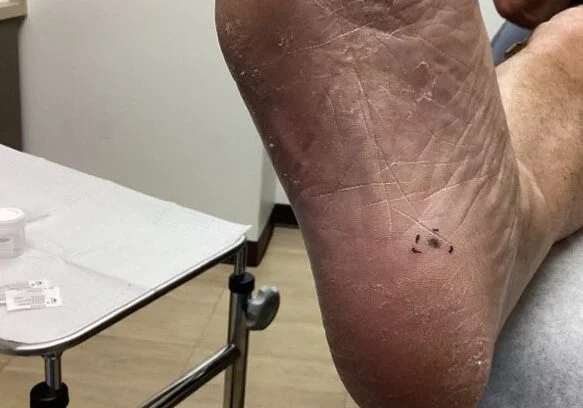
Aberrant PRAME expession in acral nevus
A 70-year-old male presented with an atypical melanocytic proliferation on his right instep. He had no personal or family history of melanoma. The clinical diagnosis was acral nevus - rule out atypia.
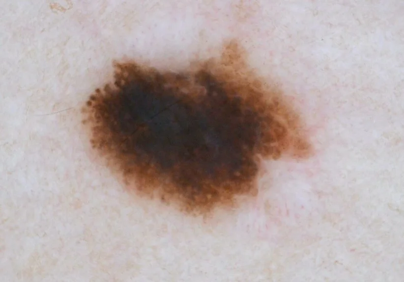
Abnormally pigmented lesion with uncertain malignant potential
A woman in her 30's presents to her dermatologist for evaluation of a pigmented lesion on her inner thigh.
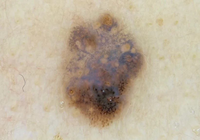
Asymmetrical Pigmented Lesion with High Clinical Suspicion for Melanoma
A 27-year-old female patient was seen by a primary care physician who recommended follow-up with a dermatologist for assessment of a concerning melanocytic lesion.
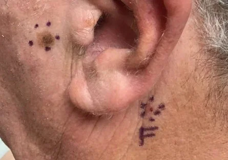
Atypical melanocytic proliferation on sensitive preauricular cheek
A 65-year-old male presented with an atypical melanocytic proliferation near the left ear. Four sites of basal cell carcinoma and seborrheic keratosis were taken at the same visit.
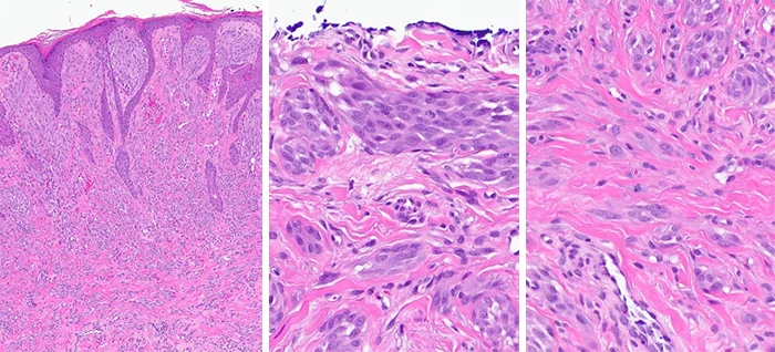
Atypical Spitz nevus versus Spitz melanoma
A 24-year-old male presents with a lesion on his left superior helix
Concerning melanocytic lesion in patient with personal history of melanoma
A 49-year-old male was seen by a dermatologist for a concerning nevus on the right superior lateral neck. The patient's age, extensive solar damage, and history of metastatic melanoma was a cause for concern.
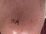
High-risk melanoma diagnosed and risk-stratified with MyPath Melanoma and DecisionDx-Melanoma
A 74-year old male wih a dome-shaped red papule on right central frontal scalp
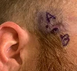
Inconclusive PRAME staining on high-stakes lesion on face
A 34-year-old male with a positive family history of melanoma presents with a lesion on the right lateral malar cheek. It was questionable if the lesion had been previously biopsied
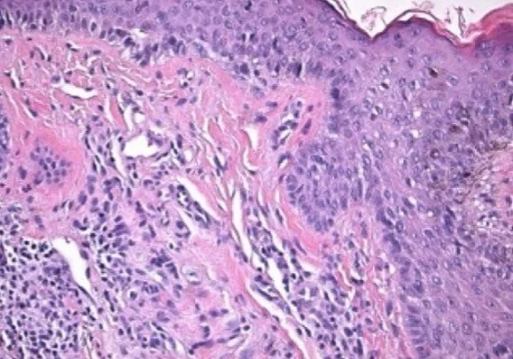
Irregular papule with atypical histopathological findings and borderline/uncertain diagnosis
A 43-year-old female patient presents to a dermatologist with a suspicious irregular multi-colored papule on the right dorsal foot.
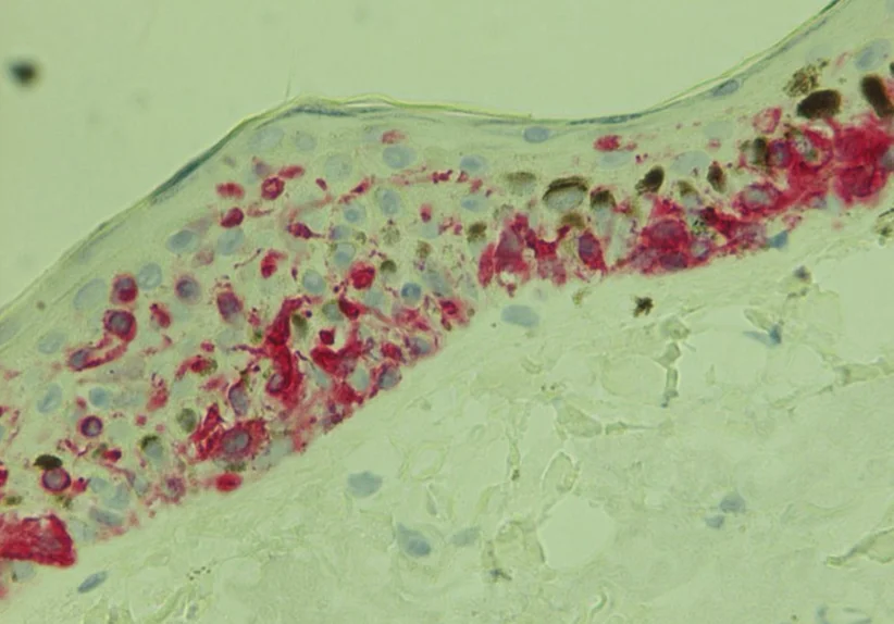
Multi-colored lesion with uncertain malignant potential
A 80-year-old male presents with a multi-colored lesion on his back. He has a previous history of extensive sun damage and non-melanoma skin cancers.
Get started with MyPath Melanoma