Case Study

Asymmetrical Pigmented Lesion with High Clinical Suspicion for Melanoma
A 27-year-old female patient was seen by a primary care physician who recommended follow-up with a dermatologist for assessment of a concerning melanocytic lesion.
Case details

A 27-year-old female presented with a 6 mm asymmetrical lesion on the back. Blue color and round structures were visualized by dermoscopy and confocal microscopy. The patient has a family history of melanoma and pancreatic cancer. Clinical differential diagnosis included melanoma in-situ, malignant melanoma, or regression of atypical nevus.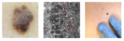

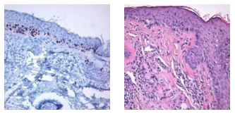
It was noted in the pathology report that there were suspicious, atypical findings including focal confluence of nests, areas of irregular epidermal distribution, and dense inflammation. A final diagnosis of melanocytic nevus, compound type, with atypical features, dense inflammation, and fibrosis was rendered by the dermatopathologist with recommendation for clinical follow-up.

Clinical findings were highly suspicious for malignancy, but pathology was not diagnostic for melanoma. The inconsistency between clinical and pathological findings left the treatment plan in question. MyPath Melanoma was ordered and resulted in a gene expression profile suggestive of a malignant neoplasm.
Following the MyPath Melanoma test result, the treatment plan was modified to include re-excision and quarterly follow-up. An addendum to the pathology report was issued noting that while not definitive, the histopathological findings could not exclude melanoma in-situ arising within a nevus.
A follow-up skin check at four months revealed an additional melanoma and two evolving pigmented nevi under close monitoring with dermoscopy.