Case Study

Flat atypical nevus on the cheek
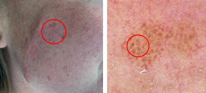
A 45-year-old female presents with a 3 mm flat atypical nevus on the right cheek.
Case details

A 45-year-old female presents with a 3 mm flat atypical nevus on the right cheek. Shave biopsy performed. Dermoscopy reveals an atypical network and perifollicular hyperpigmentation.

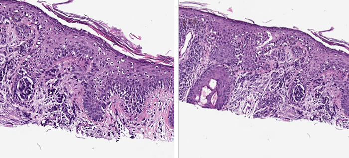
Melanocytic nevus, compound type, with atypical features.
Note: Despite a dermal cell population suggesting a congentical melanocytic nevus, confluent and irregular epidermal melanocytic involvement is an atypical finding. The nevus extends to the surgical margins and conservative re-excision is recommended.

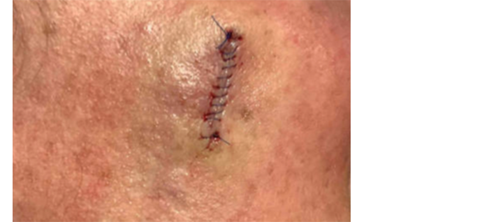
With atypia noted both dermoscopically and histopathologically, the lesion needs to be re-excised. However, due to the location of the lesion on the face, the extent of the surgical margins have a strong impact on the patient and need to be aligned with the malignant potential of the lesion.
MyPath Melanoma gene expression profile resulted in a score of 4.1 which is suggestive of a malignant neoplasm.
By combining the clinical and pathological findings with a malignant gene expression profile, the lesion was treated more aggressively and excised with 5 mm margins, consistent with a treatment protocol for melanoma in-situ.