
Clinical Summary

Archives of Dermatological Research
March 2023
Routine imaging guided by a 31‑gene expression profile assay results in earlier detection of melanoma with decreased metastatic tumor burden compared to patients without surveillance imaging studies
REFERENCE
Dhillon S, Duarte-Bateman D, Fowler G, et al. Routine imaging guided by a 31‑gene expression profile assay results in earlier detection of melanoma with decreased metastatic tumor burden compared to patients without surveillance imaging studies. Archives of Dermatological Research. 2023. https://doi.org/10.1007/s00403-023-02613-6
Results
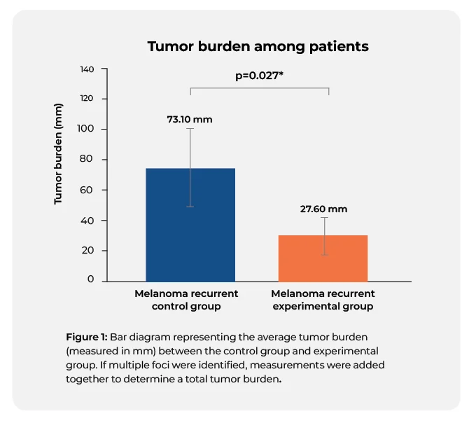
- The average tumor burden among recurrent melanoma patients in the experimental group was 27.60 mm compared to 73.10 mm in recurrent melanoma patients from the control group.
- Melanoma recurrence was detected an average of 9.85 months earlier in the experimental high-risk group, with an average detection at 25.50 months compared to 35.35 months in the control group.
- Patients with high-risk 31-GEP results who received routine imaging also showed a statistically significant improved overall survival: 76.3% versus 50.0%, p = 0.027.
Want to learn more about
DecisionDx-Melanoma?


