Spatialomics and the TissueCypher approach to Barrett’s esophagus
Emerging spatialomics techniques are enabling the spatial context of cells and tissue systems to be preserved while investigating the morphology, multiple cellular measures, expression of multiple biomarkers and spatial relationships within various cell populations and tissues. Complex molecular and cellular signatures that are characteristic of normal or pathologic states can be analyzed and contrasted in the context of cellular and tissue architecture to obtain a better understanding of the hallmarks of disease and progression, as well as the heterogeneity of the tissue microenvironment.
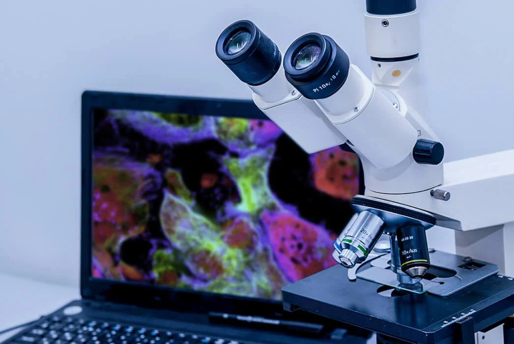
The goal of emerging spatialomics techniques in cancer is to move towards a more personalized approach for the management of patients with cancer and preneoplastic conditions. In recent years, there has been a dramatic increase in research utilizing highly multiplexed fluorescence imaging and artificial intelligence-driven platforms to analyze digital slides of patient specimens. The [.underline-move-left]TissueCypher platform[.underline-move-left] utilizes fluorescence-based spatialomics to decipher location-dependent protein expression information for patients with Barrett’s esophagus (BE). This spatialomic information is then interpreted using artificial intelligence approaches to predict the likelihood of progression to high-grade dysplasia and/or esophageal cancer in patients with non-dysplastic BE, indefinite dysplasia or low-grade dysplasia.


.webp)
