

The goal of a Barrett’s esophagus biopsy is to confirm suspected Barrett’s esophagus and to determine if any cells are growing abnormally (dysplasia). If a biopsy shows that there may be abnormal cell growth, a second expert pathologist may need to confirm the grade of dysplasia. A biopsy may also be assessed by TissueCypher for an independent prediction of cancer progression risk based on molecular biomarkers that are missed by traditional risk assessment. An individual’s risk of cancer progression informs the subsequent management and treatment of Barrett’s esophagus.
Learn more about Barrett’s esophaguscancer risk and dysplasia grading.
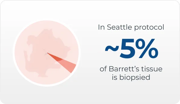
Seattle protocol for biopsy collection
The current method of biopsy collection for Barrett’s esophagus uses the Seattle protocol. This method requires a clinician to take four biopsy samples for every 1-2 cm of Barrett’s esophagus tissue – to increase the chances of detecting dysplasia. However, because the Seattle protocol biopsies only a small percentage (~5%) of Barrett's tissue, it is vulnerable to sampling and human error.
Biopsy analysis
A pathologist examines biopsies of esophageal tissue taken during endoscopic screening to confirm a diagnosis of Barrett’s esophagus and to look for abnormal or dysplastic tissue. The pathologist will view the biopsies under a microscope and grade the degree of dysplasia they see. The presence and severity of dysplasia is associated with an increased risk of progression to cancer. The physician uses this diagnosis in conjunction with the patient's other risk factors to determine the proper management and treatment.

Advancements in biopsy analysis
Extracting meaningful molecular, cellular, and morphological information from biopsies obtained during an upper endoscopy provides an additional source of information that can inform clinical decision-making. molecular changes in Barrett’s esophagus tissue that indicate an elevated risk of progression to cancer can precede signs of dysplasia, and can also extend out from an area of dysplasia creating a “field effect” around dysplastic tissue.
TissueCypher is an AI-driven precision medicine test developed to analyze these molecular changes in Barrett’s esophagus biopsies. A recent pooled analysis by the Mayo Clinic found that TissueCypher’s risk assessment was an independent predictor of progression to high-grade dysplasia or cancer, and was a stronger predictor than any other risk factor
Iyer et al., 2022
In another study, GAPP2, TissueCypher was able to identify 83% of high-risk patients that were missed by the current standard of care (Critchley-Thorne et al., 2017).
Find out more about TissueCypher


