Case Study

Inconclusive PRAME staining on high-stakes lesion on face
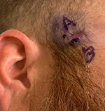
A 34-year-old male with a positive family history of melanoma presents with a lesion on the right lateral malar cheek. It was questionable if the lesion had been previously biopsied.
Case details

A 34-year-old male with a positive family history of melanoma presents with a lesion on the right lateral malar cheek. It was questionable if the lesion had been previously biopsied. Given the heterogeneous appearance, two biopsies were collected.

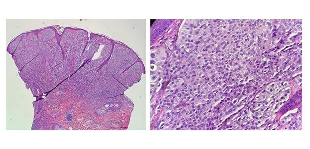
Sections reveal a proliferation of melanocytes in the dermis that show some unusual features, mostly in the upper half of the lesion. Features include large pleomorphic melanocytes with large inclusions/pseudoinclusions, dusky abundant cytoplasm and some spitzoid features. However, at the base of the lesion there appears to be some maturation of the melanocytes and less atypia.
SOX10 highlighted the melanocytes while PRAME showed diffuse although patchy/weak staining in the melanocytes. Notably, PRAME was stronger staining in the more superficial melanocytes that showed abundant pleomorphism while weaker staining in the deeper melanocytes. HMB45 showed patchy staining throughout but was mostly positive in the larger more pleomorphic melanocytes. Ki-67 highlighted the more superficial melanocytes at a rate of 5-7% while the deeper melanocytes were mostly negative. Therefore, this is highly worrisome for a melanoma arising within an intradermal nevus.
Preliminary diagnosis: Mostly intradermal melanocytic proliferation with unusual features

While the lesion was highly worrisome for melanoma, PRAME staining was inconclusive. Given the high stakes of diagnosing a deep melanoma on the face of a young person, GEP testing was ordered to objectively classify the malignant potential of the lesion.
MyPath Melanoma gene expression profile testing for the two biopsy samples resulted in scores of 2.1 and 6.4 which are both suggestive of a malignant neoplasm.
Malignant melanoma, right superior lateral and right lateral malar cheek, 2.5 mm depth
NOTE: Histologic sections reveal the malignant melanocytes reach into the reticular dermis, making this a Clark's level IV. There is 1 mitotic figure identified in part A in the superficial papillary dermis and no ulceration appreciated. There are no features of regression, but there are some tumor infiltrating lymphocytes. Final pathologic stage: pT3a (AJCC, 8th Edition, 2017).
Patient treatment plan: Patient was referred to oncology specialist for a systemic work-up to include imaging, consideration of SLNB, and presumably a WLE. Outside institution confirmed melanoma diagnosis.