Dr. Daniel Tseng: Case 1

TissueCypher reveals a missing critical detail

A 69-year old male Barrett’s esophagus (BE) patient arrived at Dr. Tseng’s practice after moving to Oregon from another state. The patient was anxious about his BE, but that concern is common for many BE patients who know that they have a precancerous condition and feel uncertain about their risk of developing cancer.

Read the case details below.

Endoscopic findings

Dr. Tseng performed an upper endoscopy and collected biopsies, revealing a Prague classification of C3M4, no hiatal hernia, and a diagnosis of non-dysplastic Barrett’s esophagus. Traditional – yet subjective – management based on these findings would be surveillance at a one-year interval. 

Lower esophagus: 
  • BE segment length: Prague C3M4 
  • No hiatal hernia
  • Pathology returned non-dysplastic (NDBE)
Traditional Management: 
  • Low risk clinical profile due to NDBE
  • Treatment recommendation: 1 yr. surveillance due to anxiety and long segment

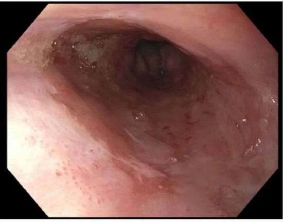
Endoscopy image from lower third of the esophagus showing salmon-colored mucosa and no visible lesions

Endoscopy image from lower third of the esophagus showing salmon-colored mucosa and no visible lesions

TissueCypher results

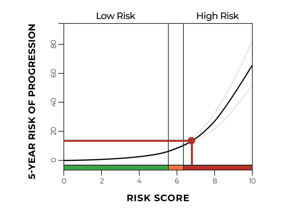
When the TissueCypher results arrived, Dr. Tseng was concerned by the high-risk score and 14% probability of progression over 5 years. That prompted Dr. Tseng to have a discussion with the patient during which the prior diagnosis of EAC was revealed, and the patient agreed to move forward with radio-frequency ablation to remove his BE. 

  • Risk class: High
  • Risk score: 6.7
  • 5-year risk of progression: 14% 

TissueCypher-guided management

  • Change in management: Upstaged

"Going back through this patient's history really validated the value of TissueCypher. Even though he had a very bland-appearing Barrett's segment... he actually had a high-risk of developing esophageal cancer." 

Daniel Tseng, MD, FACS