Abnormally pigmented lesion with uncertain malignant potential
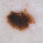
A woman in her 30's presents to her dermatologist for evaluation of a pigmented lesion on her inner thigh.
Case details

A woman in her 30's presents with an isolated lesion with irregular pigmentation. When evaluated by dermoscopy the lesion showed a global disorganized pattern including focal pseudopods and a central eccentric dark structureless area. Reflectance confocal microscopy showed dendritic and pagetoid cells in the epidermis. There is also an atypical ring pattern with cells infiltrating the rete and junctional thickening.
Overall these findings are suggestive of melanoma. A narrow excisional biopsy with only 1mm margin was obtained to sample the lesion in its entirety. Clinical differential diagnosis includes melanoma or spitz nevus.

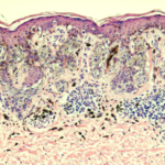
Single and aggregated atypical spindled and epithelioid melanocytes at the dermal-epidermal junction with confluence of nests. Occasional pagetoid melanocytes are noted. There is lymphohistiocytic inflammation with melanophages in the dermis. The diagnosis was rendered as a melanocytic neoplasm with usual features.
Note: The differential diagnosis is between that of a Clark's nevus with spitzoid features or a very subtle melanoma measuring approximately 0.25 mm in Breslow thickness. The margins are free of neoplasm. Former diagnosis favored.

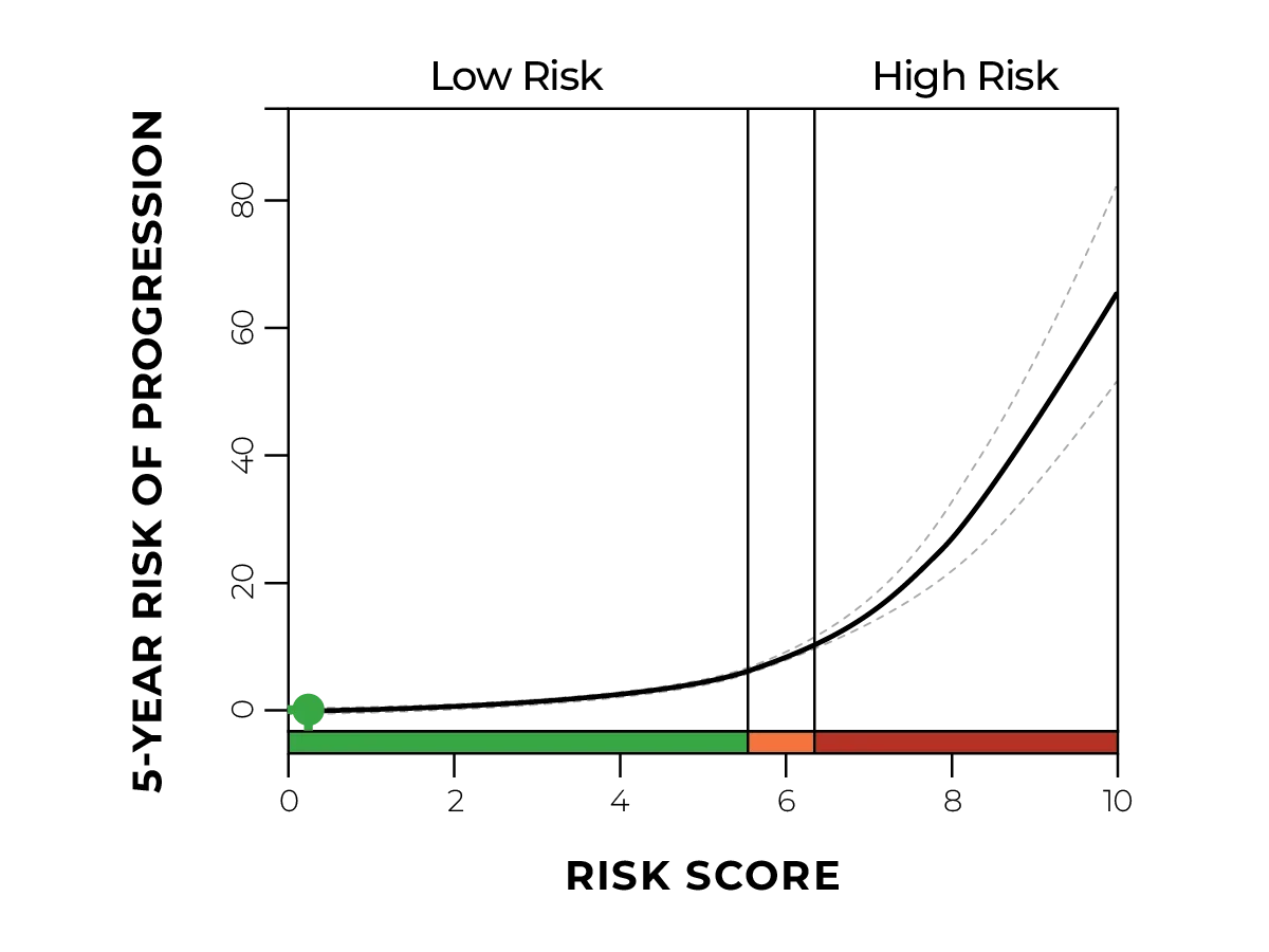
Due to equivocal histopathology and diagnosis with uncertainty in the malignant potential of the lesion, the pathologist requested both a second opinion consultation and diagnostic gene expression profiling (GEP). MyPath Melanoma resulted in a gene expression profile suggestive of a benign neoplasm.

By incorporating both the benign GEP test result and an expert consultation (reported as benign compound Spitz nevus), the dermatopathologist adjusted the preliminary diagnosis to reflect a benign melanocytic neoplasm. No re-excision was performed, the patient did not receive a diagnosis of invasive melanoma and she was managed with routine follow-up.

If melanoma had not been ruled out, the patient may have been treated with a wide local excision with 1 cm margins and follow-up intervals for invasive malignant melanoma.