Atypical melanocytic proliferation on sensitive preauricular cheek
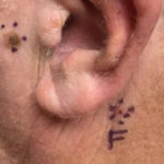
A 65-year-old male presented with an atypical melanocytic proliferation near the left ear. Four sites of basal cell carcinoma and seborrheic keratosis were taken at the same visit.
Case details

A 65-year-old male patient was referred to dermatology for an atypical melanocytic proliferation near the left ear. The lesion was 7 mm in diameter, on left superior preauricular cheek. It was a reticulated, light tan macule, changing size and color. Clinical differential diagnosis included lentigo, lentigo maligna, and melanoma in-situ.

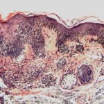
The H&E staining showed cohesive nests of small round melanocytes at the tips and bases of the rete ridges. An increased number of single melanocytes was noted along the dermoepidermal junction with some bridging noted. There was also apparent follicular extension of the junctional melanocytic proliferation as well as scattered pagetoid melanocytes appreciated in the mid to upper layers of the epidermis.
Melanoma could not be ruled out by H&E alone. Treatment recommendation not provided.

There was a lingering concern over the possibility of melanoma in-situ and the inability to rule out melanoma by histopathology alone. Due to the sensitive location of the lesion, testing was ordered to guide the surgical margins.
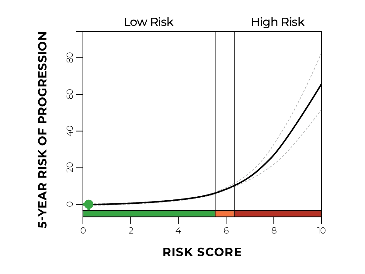
MyPath Melanoma resulted in a gene expression profile suggestive of a malignant neoplasm.

The test results led to a more confident diagnosis of melanoma in-situ arising in a dysplastic nevus and the dermatologist was able to adjust the surgical margins accordingly.

When it isn't possible to rule out a melanoma diagnosis through the pathology work-up alone, diagnostic GEP testing can provide additional information to obtain a more confident diagnosis and help guide the surgical margins.