Multi-colored lesion with uncertain malignant potential
A 80-year-old male presents with a multi-colored lesion on his back. He has a previous history of extensive sun damage and non-melanoma skin cancers.
Case details

Dermoscopy showed atypical network with grey dots and granules. Structures were predominant at the periphery with structureless areas in the center. Confocal microscopy demonstrated atypical cobblestone pattern in the epidermis with pagetoid cells. Loss of typical meshwork pattern in the dermal-epidermal junction. Bright cells infiltrating the rete and numerous spindle shaped structures. Clinical differential diagnosis included melanoma on sun-damaged skin or evolving melanoma in-situ.


A comprehensive histopathological work-up included immuno-histochemical staining. Melan-A and S-100 confirmed the diagnosis of a nevus and Melan-A showed a confluence of melanocytes at and above the dermal-epidermal junction. Lentiginous epidermal hyperplasia and hyperpigmentation was noted. At the dermal-epidermal junction, discohesive nests of melanocytes and atypical melanocytic hyperplasia were identified. Superficial perivascular infiltrate with melanophages was identified in the underlying dermis.
A final diagnosis of Clark's nevus with unusual features was rendered with a recommendation to re-excise.


Given the conflicting findings between the clinical impression (melanoma) and diagnosis (Clark's nevus with recommendation to re-excise), the malignant potential of the lesion was uncertain.
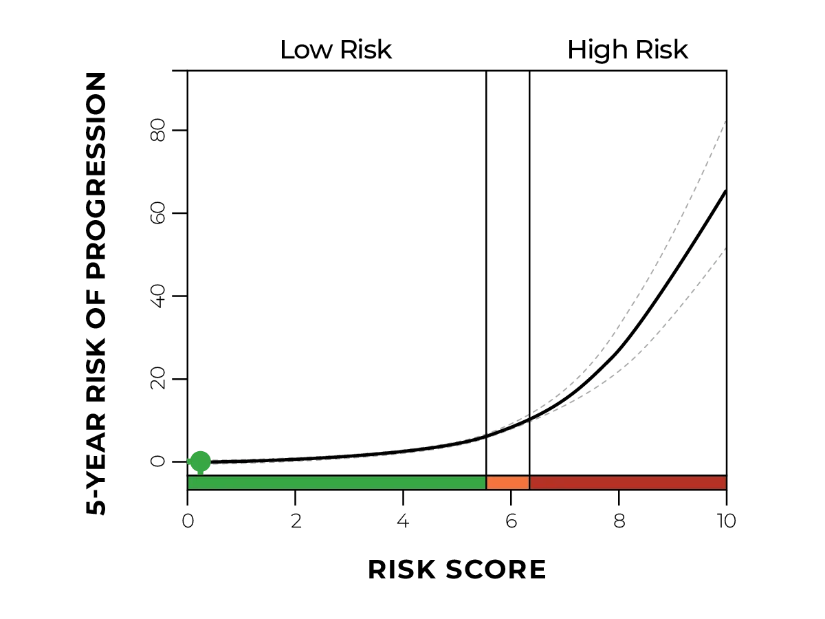
DiffDx-Melanoma resulted in a gene expression profile suggestive of a malignant neoplasm.

Due to the DiffDx-Melanoma test results, the dermatologist confirmed their clinical suspicion and was able to proceed with surgical excision using margins consistent with a melanoma in-situ. The patient management was adjusted to reflect more frequent follow-up appointments.

DiffDx-Melanoma GEP testing can be used to confirm or deny suspicion of a lesion with unknown malignant potential. The additional information provided can help ensure that the patient has the correct treatment and management plan.