Irregular papule with atypical histopathological findings and borderline/uncertain diagnosis
A 43-year-old female patient presents to a dermatologist with a suspicious irregular multi-colored papule on the right dorsal foot.
Case details

The patient presented with a 7 mm in diameter, irregular, multi-colored raised papule on the right dorsal foot. Clinical differential diagnosis included melanoma or an irritated nevus.

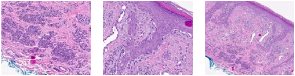
Lesion showed compound proliferation of melanocytes. In the epidermis, there were also many single melanocytes with limited pagetoid scatter. In the dermis, there were many nests, cords, and small syncytia with maturation or pseudo-maturation. Scattered inflammation was apparent. PRAME expression was absent. Due to the difficulty of the case and suspicious features, outside consultation was requested. p16 staining was performed and was diffusely positive. Neither PRAME nor p16 staining demonstrated biphasic reactivity as can be seen in the setting of melanoma. The consulting pathologist's impression was that the lesion was an extreme compound acral melanocytic nevus. No treatment recommendation was provided.

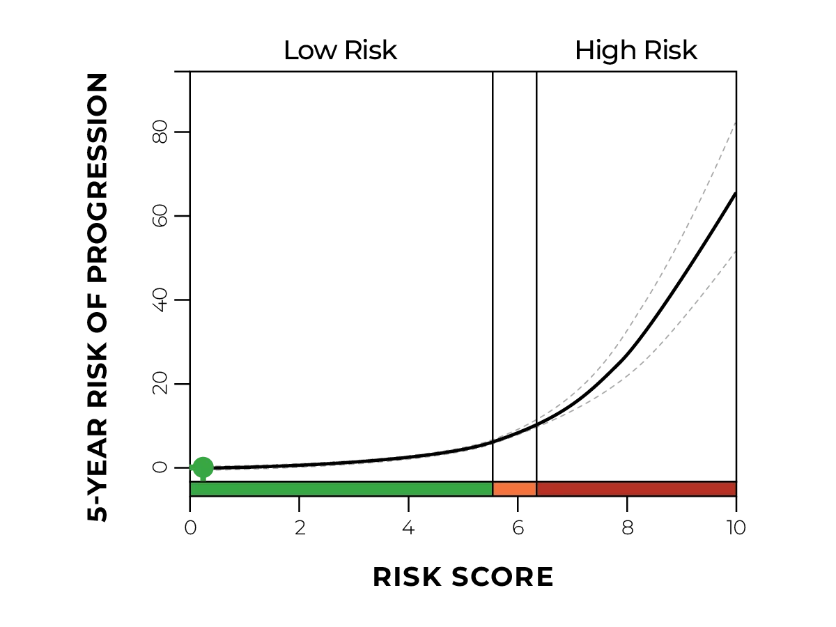
Even after an extensive workup with additional ancillary testing and a second opinion consultation, the diagnosis was still borderline or uncertain and diagnostic GEP was ordered to help eliminate this uncertainty.
DiffDx-Melanoma resulted in a gene expression profile consistent with a benign neoplasm.

By incorporating the results of the IHC stains, second opinion consultation, and DiffDx-Melanoma GEP results, a final diagnosis of atypical acral nevus was rendered. Given the benign diagnosis, the margins on the surgical excision were narrowed to 0.5 cm.

DiffDx-Melanoma GEP testing provided valuable objective information to both the dermatopathologist and dermatologist, confirming the diagnosis and helping to guide patient management decisions. The benign diagnosis narrowed the excision margins and allowed for less frequent follow-up appointments.